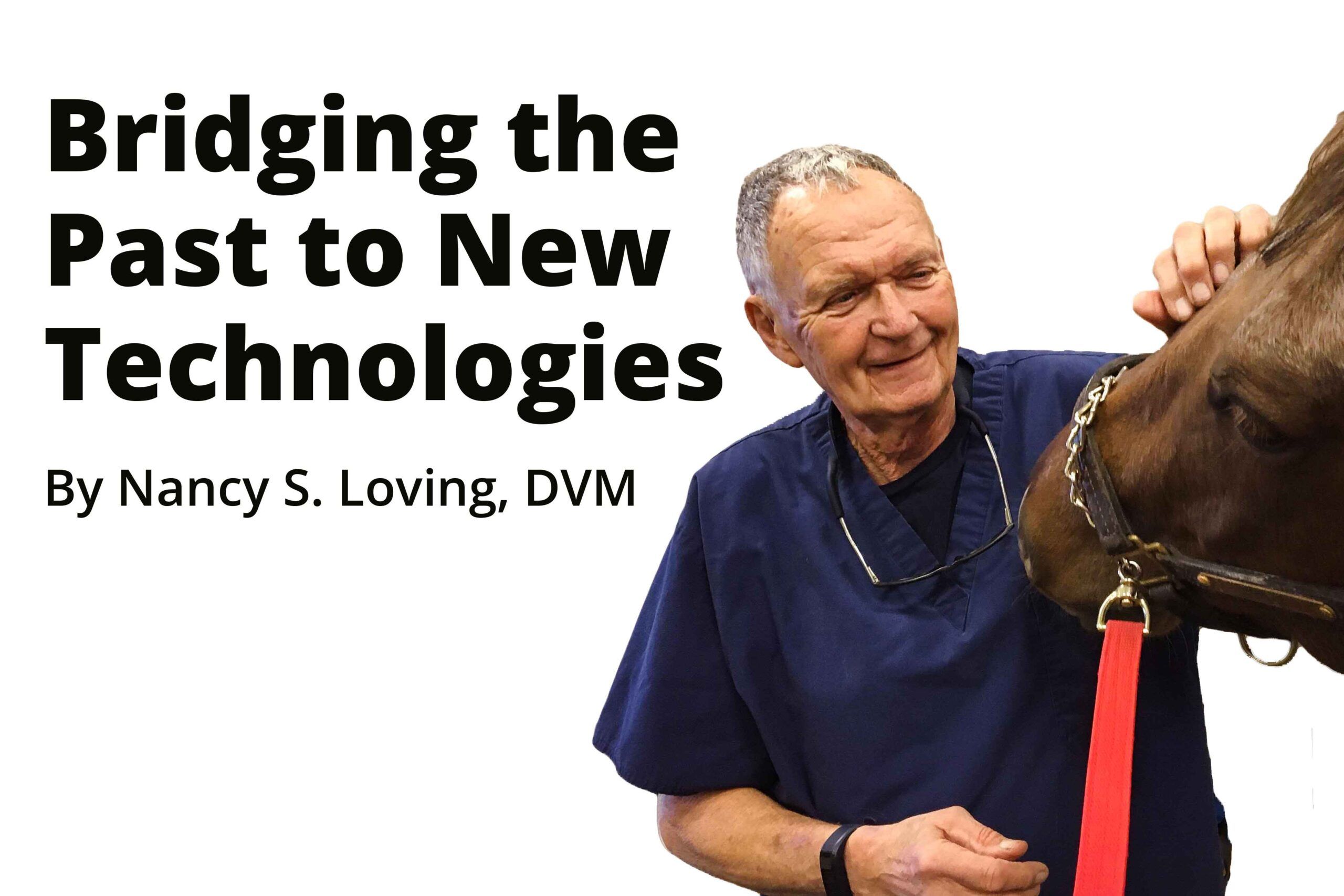
Featuring Dr. Ron Genovese – Insight from 56 Years in the Practice of Lameness Diagnosis
By Nancy S. Loving, DVM
There have been some giants in the equine veterinary industry who have paved the way for following generations of veterinary professionals. In a recent interview, one of those pioneers — Dr. Ron Genovese — graciously shared his insights from 56 years in the practice of lameness diagnosis.
His story is an example of how new techniques and technology can combine with a lifetime of veterinary experience to improve a practitioner’s skill and ultimately lead to products as novel as The Equinosis Q with Lameness Locator developed by Dr. Kevin Keegan.
Genovese grew up riding Thoroughbred racehorses, so he naturally gravitated to the racetrack following veterinary school at the University of Pennsylvania. In those days, the available tools a practitioner had were a stethoscope, thermometer, hoof testers, hands-on ability to palpate and flex a limb, and access to internal views generated by an x-ray machine. Most seasoned track practitioners only looked at horses in the stall but Genovese’s boss, Joe Solomon, DVM, was progressive and wanted to trot the horses out. Also, in those days half a century ago, whatever the trainer at the racetrack said was accepted as gospel by the horse owner. The veterinarian was very much at the beck and call and whims of the trainer because there was little opportunity to contact the racehorse’s owner.
Rather than trotting the horse to determine which leg(s) was a problem, most practitioners took the trainer’s information at face value, and a brief hands-on evaluation would point the veterinarian in a direction of therapy. Yet, Genovese was convinced there was a better way. His veterinary training at New Bolton was inspired by some great practitioners who emphasized that an examination included taking a horse out of the stall, trotting it on straight lines and circles, careful palpation, flexion testing, and radiographic images. At the time, many racetrack owners and veterinarians were opposed to this more active method of diagnosis, saying there simply wasn’t time to move the horse around and get through the day’s case load.
To help train his ear to a cadence of asymmetric footfalls, Genovese felt trot-outs were important in addition to their value as a visual aid to distinguish lameness. He says, “I was motivated to assess the patient as thoroughly as possible, identify the problem, and then implement long-term rehabilitation.” This approach was a considerable forward step in lameness diagnosis and management, quite different from what was being done by other veterinary practitioners at the time.
Sharing of experiences by equine veterinary colleagues including those who influenced Genovese contributed to the inception of the American Association of Equine Practitioners (AAEP). The motivation to disseminate information to equine practitioners was part of the germination of the Proceedings book put out annually by AAEP.
Another challenge for Genovese during his formative years in veterinary medicine was that as a young veterinary professional, trainers didn’t take him seriously and wouldn’t hesitate to change his diagnosis. “This was frustrating because I didn’t get the experience of mistakes,” he mused.
Because the racetrack trainer dictated most medical services, Genovese worked out a compromise with his clients where he would give ground for “x” number of things as long as they would allow him to trot out the horse, palpate, flex, and radiograph. By 1978, he had developed a good reputation on the track along with acceptance by the trainers. Finally, he could achieve the experience of both mistakes and successes.
Genovese describes a catastrophic occurrence in 1980 that affected him — It was a trigger point. A horse entered in the Ohio Derby was galloped in preparation for the race and developed a warm ankle. The horse was examined, trotted, and radiographed with negative findings. He was raced with a devastating result: Severe injury to the suspensory apparatus that resulted in euthanasia. At that time, there was no known way to image ligaments or tendons. Genovese queried, “Why not identify a problem prior to a race?”
Sometimes the stars align just right to bring people together at the right time and yet another contemporary pioneer entered the scene — Dr. Norman Rantanen who was equally motivated to care for a horse’s welfare. At that time, Rantanen had credibility in the equine veterinary world, and was fascinated with new technology: Ultrasound.
Still, there was pushback to this novel ultrasound imaging technique. Curious veterinarians who came to look at the equipment would wonder how the machine could possibly do more than what their professional fingers could tell. We are well aware of how this turned out — ultrasound is a commonplace and critical tool in lameness diagnostics in the 21st century.
Early on, Genovese liked the concept of ultrasound and its credible source through Dr. Rantanen, so he transitioned from solely racetrack practice to include sports medicine horse care. At the racetrack, nerve blocking as a diagnostic tool was not performed due to conflicts with drug testing, and also because not enough owners and trainers were motivated to “preserve” horses. Upcoming technology like ultrasound, nuclear scans, MRI, and eventually CT scans were more expensive diagnostic tools than track owners wanted to pursue – it took many years for advanced technology like this to become an accepted standard of care.
Still, Genovese desired to look deeper into a horse’s condition with the recognition that lameness is a sign and there was a need to get to the root cause and a potential strategy to heal a problem. As he transitioned more completely into sport horse care and away from the track experience, he contemplated how to obtain the most information possible about a horse’s condition. He wondered, “Who is the ultimate diagnostician of each horse?” He came to the conclusion that it is the rider, especially for sport horse equine athletes. These riders can sense when something feels “off” although the naked eye can’t always see a problem. He notes, “Most sport horses experience occupational or bridle lameness, which tend to be subtle issues.” When caught early before becoming a full-blown problem, steps can be taken to achieve resolution.
As time progressed, more advanced technology became available in equine practice: Digital x-ray, digital ultrasound, nuclear scan, MRI, arthroscopic surgery, shockwave, thermography, advanced medications like hyaluronic acid. Another pioneering giant in the equine field, Dr. Kevin Keegan, was on a quest for a method to achieve objective analysis of lameness. His research efforts into kinematics and lameness led to development of The Equinosis Q with Lameness Locator. Genovese stresses that equipment such as ultrasound and The Equinosis Q are invaluable adjuncts to a practitioner’s clinical skills and improves on what the hands can feel, and the eyes can see. Genovese discovered that The Equinosis Q was a technological tool that could identify and corroborate subtle lameness issues of which a rider might be aware, but a veterinarian could not identify through subjective examination. For the last 10 years of his practice life, he used The Equinosis Q on every single horse he evaluated. He stresses how much it taught him personally in addition to serving as an invaluable tool to help teach his students how to “see” gait asymmetries.
The Equinosis Q has served a great function for Genovese as a simple yet effective tool to incorporate as a part of his lameness workups. He says, “As a proven diagnostic tool, it quantitates gait asymmetry and serves as a backup and support of my clinical assessment. It also helps to train students and young veterinarians and develops their confidence in lameness diagnosis. This enables older practitioners to spread the work load to their less experienced associates.”
In addition, he advocates that it is very helpful to use before and following diagnostic nerve blocks to remove any subjective “desire” for results. Not only does The Equinosis Q eliminate subjective impressions from the evaluation, but he also discovered that owners appreciate such objective documentation results. Combining objective documentation with a thorough clinical workup is good for successful sleuthing of a lameness issue.
Another useful means of incorporating The Equinosis Q into lameness exams is that besides supporting clinical impressions, Genovese stresses that it becomes an easily-inserted part of the medical record. “Documentation of a horse’s gait in this objective manner improves the integrity and reputation of the practitioner,” reports Genovese. In fact, he notes that for a follow-up exam, it is possible to send out a technician or associate veterinarian to gather follow-up data and this frees up the older practitioner to provide more complicated services. The Equinosis Q then tracks how a patient is doing through time following appropriate therapy or once off medication.
In addition, data from The Equinosis Q provides documented information for follow up exams because as time passes, it is not always easy for a practitioner to remember subtleties of every case. With his case load of 7-12 lameness exams each day for six days a week, it would be extremely challenging to remember every little detail of previous exams on every horse. The objective report is there as certain confirmation of how the horse appeared at a previous time period, and it also allows a new set of veterinarian eyes to have definitive historical data when examining a horse for the first time.
He recounted a story about a stem cell study that required confirmation of The Equinosis Q with a visual exam. In the follow-up study, despite the fact that three horses were deemed sound with visual inspection, The Equinosis Q still indicated gait asymmetry. For Genovese, this was insightful information that helped inform rehabilitation decisions on when to progress a horse into more work.
As an additional strategy, Genovese suggests that The Equinosis Q may be valuable for obtaining a baseline on each horse in clinical practice. This can then be followed up once or twice a year at a Wellness check to help identify beginning or subtle lameness concerns. The only downside he can report is that this takes time if one is unfamiliar with using the technology; however, one charges for the service, so it is part of practice revenue.
Genovese recognizes that for some, initial use of The Equinosis Q may be intimidating. With practice, the sensors can be quickly mounted on the horse (even without assistance from a technician) and then the typical lameness exam proceeds with lunging or riding, flexion tests and diagnostic nerve blocks. The inertial sensors report information to the computer tablet in real time, and it is all there instantly for analysis.
At his innovative equine practice, Dr. Genovese has been diligent in implementing new technology throughout his 56 years in equine practice. He has seen the industry evolve to the highly technical and professional level that we experience today. His personal commitment to pursue and implement the scientific method and evidence-based medicine has earned him a great reputation and respect in the equine veterinary industry, and he has played a role in influencing and mentoring following generations of equine practitioners. He likes to think that he has been a bridge from the past to the technological advances of today. We have all benefitted from his pioneering spirit that has helped elevate lameness diagnostic methods throughout the decades.
***


Leave a Reply