Motion Sensor Technology: Advancing Racehorse Safety One Step at a Time
LEXINGTON, KY – The Sixth Annual Tex Cauthen Farrier, Veterinarian and Researcher Seminar was held on January 23, 2022 at the Maxwell H. Gluck Equine Research Center on the University of Kentucky campus. Focused on “Protecting the horse, the hoof and the biomechanics of the hind limb,” the day-long seminar was open to both in-person and online attendees.
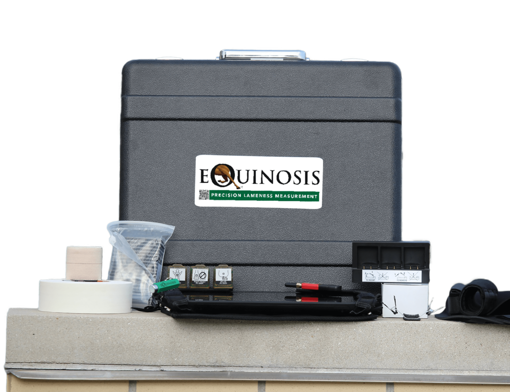
Equinosis Q inventor Dr. Kevin Keegan, in his third year speaking at the seminar, provided an update on the development of the tool and studies, reiterating why the technology has become increasingly important as pressure mounts on the racing industry to provide increased safety measures.
Keegan covered the evolution of the Equinosis Q and what movement parameters should be measured for repeatable assessment of lameness. After investigating many measurements, including limb swing, stride length, joint angle changes and others, vertical displacement of the head and pelvis on the midline of the horse was determined to be the most sensitive and accurate.
Keegan noted that lameness is best detected at the trot; studies completed at the canter and gallop did not yield reliable detection of lameness. “It seems that at the trot, the horse has many mechanisms to shift weight around due to pain, and this can be picked up,” Keegan explained. “When the horse is galloping, it is forced into a particular movement that is difficult to change.”
The next challenge facing mainstream acceptance of inertial sensors is to help non-users understand that although sound horses are often quite symmetrical in their measurements, asymmetry (measurable lameness) does not necessarily indicate pathology – no matter how counter intuitive that may seem to some. Keegan said that there are currently no recognized reference ranges for objective measurements of lameness among Thoroughbred racehorses; but the best way to obtain these would be the continued evaluation using inertial sensors. Gathering data from a large number of horses on a variety of tracks, and then following them throughout their racing careers, will allow veterinarians to better understand what amount of asymmetry is “normal” and whether an individual horse has what is considered a “usual” pattern of asymmetry.
To highlight the varying degrees of asymmetry measured on actively training racehorses, Keegan reviewed data from a pilot study of 73 horses over 16 weeks of racing at Hollywood Gaming at Mahoning Valley Race Course and at JACK Thistledown Racino in Ohio, and of 36 horses over 18 weeks of racing at Prairie Meadows Casino, Racetrack and Hotel in Iowa. Data was collected at least five times on 79 of the horses.
A modified theoretical reference range for racehorses was calculated; the estimated reference range was approximately double the experimentally determined reference ranges for forelimb and hindlimb lameness in a broad population of horses, most non-racehorses.
Data extrapolated from the study showed that 29.5 percent of the Iowa horses and 50.7 percent of the Ohio horses measured outside one of these theoretical reference ranges at least once throughout the study period. Examining these results, Keegan summarized that it is common for active Thoroughbred racehorses to measure with some degree of lameness, at least intermittently, without necessarily requiring intervention. It is still unknown if lameness correlates to catastrophic breakdowns.
Keegan shared a few examples of horses from the study. One example was the only documented catastrophic breakdown, which occurred after the study concluded. The horse had a diagonal right front and left hind lameness, measured intermittently over the 16 weeks of the study. It raced three additional times over 2.5 months following the conclusion of the study, when it sustained a biaxial sesamoid bone fracture in its left front, the opposite forelimb to which the horse had measured with lameness previously.
Keegan went on to offer another example of a horse with a similar pattern of right front lameness that went on to race 18 times without incident following the study conclusion. “So you can see, the relationship between lameness and injury is not simple,” Keegan remarked.
To conclude, Keegan does not recommend using inertial sensors just once immediately before a race as a screening tool to assist in determining which horses should be deemed “sound” enough to race. Using the machine just once could lead to a multitude of horses being excluded from racing as their unique asymmetry is not known and could be taken out of context as at-risk for catastrophic musculoskeletal injury (CMI). Keegan advised, “Evaluate the horse regularly by measurement in between racing and training days, when lameness is more likely to be expressed.” Keegan added, “You have time to evaluate what the meaning of that measurement is.” This allows for physical examination, limb evaluation and good quality imaging when necessary.
Keegan says future studies should focus on patterns of lameness associated with CMI. Measuring horses multiple times between racing and training would allow for the accumulation of concrete data that can be archived, searched and shared between veterinarians and racetracks – especially needed when multiple tracks and racing jurisdictions are involved, he notes. Researchers and veterinarians would then be able to determine if there is an association between lameness and CMI.
Adoption of Inertial Sensors in Racing
Keegan cited that some veterinarians are still reluctant to use lameness measurement for a myriad of reasons. These can include:
1. Fear and pride. Some vets feel they can see lameness well enough and therefore do not need to measure it. “Even if that were true, it’s totally missing the point,” Keegan says. He emphasized, “For our profession, [measuring lameness] creates a database we can search and analyze for patterns that we may find out are useful.” It also allows for objective comparison between dates and different evaluators.
2. It’s “too difficult to get the data.” “It takes two minutes to put the sensors on,” says Keegan. And trotting takes time whether it’s the human eye or the sensors measuring it, he says. If the horse is difficult in hand, the horse can be ridden, which allows for easier collection of many strides.
3. Concern that the Equinosis Q won’t pick up bilateral lameness. “This is really a straw man argument,” Keegan says. “While it is true theoretically, it is not actually true in practice.” Bilateral lameness is harder to detect whether it is being determined visually or using motion sensors, Keegan notes. However, the horse is not usually exactly as lame in one limb as it is the other, on every stride. “This hardly ever happens,” says Keegan. “Lameness as a clinical sign is not that consistent.” It can switch between left and right over the exam, and over time. The use of inertial sensors and the evaluation of multiple strides can detect nuanced lameness more sensitively. Lameness can also be lateralized through flexions or moving the horse in circles, he says.
Highlighting the Need for Ongoing Analysis
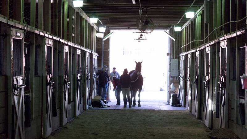
Dr. Rhodes Bell of Bell Equine Surgery and Lameness in Central Kentucky, and Dr. Clara Fenger of Equine Integrated Medicine in Georgetown, KY, presented numerous case studies highlighting the benefits of repeatedly using the Equinosis Q over a period of many months. Bell and Fenger are involved in an ongoing study in which racehorses are evaluated monthly using the Equinosis Q to see if injuries can be detected earlier. The horses will be enrolled in the study until their racing career is over or until ownership is transferred through a sale or claim.
Bell noted that there are multiple reasons a horse could be lame, from osteochondritis dissecans (OCDs) to arthritis to trauma. Currently, a primary concern in racehorse welfare is the identification of stress fractures and their potential role in catastrophic injuries. Both noted that asymmetrical movement due to developing stress fractures may be very subtle and nearly impossible to see with the naked eye.
Bell shared that a human’s ability to detect asymmetry has a threshold of about 25 percent (of the total vertical excursion). Additionally, he noted that clinicians agree with a confirmed lameness about 75 percent of the time when the lameness is up front and 67 percent of the time when the horse is lame behind. Use of inertial sensors allows the limitations of human perception to be overcome with objective data.
Dr. Fenger then offered case studies of a few of the horses she and Dr. Bell have been tracking using the Equinosis Q.
Case Study #1: Using Q Data to Guide Management Over 18 Months
The first case study Fenger presented involved a 2-year-old Thoroughbred racehorse that had a presumptive tibial stress fracture after coming in acutely lame in his right hind after a work. The gelding had four months off before an initial Equinosis Q evaluation; he was then followed over the next 18 months of his racing career. Fenger noted that the horse was not blocked immediately after his initial lameness as the potential for boisterous behavior and additional harm to the horse is of concern.
The Q was first used when the horse was about to return to training; results showed mild residual lameness, but the horse was gradually returned to training. After five months of training, the lameness worsened acutely, again arising after a work. It was suspected that the horse returned to work too quickly and that it was experiencing a recurrence of the tibial stress fracture. The horse received four months of rest, this time with monthly monitoring with the Q. After four months, the right hind lameness was no longer measurable and the horse was returned to training.
Again, the pattern repeated: Five months after resuming training, the horse came up lame in the right hind after a work. The horse was radiographed and the images showed no active remodeling of the tibia (thus the horse was in minimal danger of a catastrophic breakdown of the tibia).
The horse was then blocked to try to pinpoint the location of the lameness. Interestingly, when the horse was blocked to the right hind foot, the lameness swapped to the left hind. The cause of lameness was determined to be a negative palmar angle that placed excess strain on the impar ligament, which attaches the navicular bone to the coffin bone.
The horse’s health care team chose not to have an MRI done as the horse was relatively inexpensive. Instead, they relied on farrier Colby Tipton to determine how best to help the horse. Tipton removed the toe grabs on the hind shoes, trimmed to balance the hooves and removed the extra toe (racehorses are notorious for having long toes and low heels). By ensuring the horse didn’t go too long between shoeing cycles, the toe remained set back, reducing the ground-up impact on the horse’s body.
This corrective shoeing, plus injecting the gelding’s coffin joint, resolved the horse’s ongoing lameness issues. The horse went on to race in months 17 and 18, finishing third once. The horse has not been lame since; he continues to have monthly evaluations using the Q.
Case Study #2: Using the Q to Identify Gait Signatures
The second case study Fenger offered highlighted the utility of using measurement to determine when a horse can return to training. This case followed a 3-year-old filly that flipped in the gate during her first race on November 18, 2020, causing a traumatic fracture of the greater trochanter of the left hip.
At two months post injury, the filly was evaluated and measured with the Q, which showed a left hind pushoff lameness and right hind impact lameness. After an additional month of rest, she was re-evaluated using the Q, which showed that all hindlimb lameness had resolved. A recheck ultrasound showed that there had been smooth remodeling and the filly was cleared to return to training. Interestingly, the mild right hind impact lameness was measured repeatedly over the following year, but the left hind lameness associated with the injury never recurred. She subsequently ran five races, winning two.
Though the filly is considered “serviceably sound,” she consistently measures with a right hind asymmetry; this movement pattern has become the filly’s “gait signature”. Similar to how a person’s fingerprints are unique to them, the way a horse moves is unique to each individual.
Fenger and Bell’s take home messages are that:
- The Equinosis Q is an effective tool to determine when horses on layup for an injury can safely return to training
- A single lameness exam utilizing this tool is not effective as a screening mechanism to prevent injury
- An objective “gait signature” can be identified when multiple exams are conducted over time and the asymmetries remain the same
Highlighting the Data from Motion Sensors can Save Lives
Dr. David Lambert updated Tex Cauthen attendees on the data that the Stride Safe system can provide. Stride Safe is a tool that has was developed from the StrideMASTER, an instrument that used sensors to supply handicapping information to bettors. Tweaked in design to provide information that will protect racehorses, Stride Safe uses GPS biometric sensors as performance predictors.
“Stride Safe is not a diagnostic tool,” Lambert reiterated. “The goal is to point out animals that are particularly at risk” of catastrophic breakdowns. “This product can ‘flag’ horses, but it can’t make recommendations on what can be done with these horses [to make sure they compete safely],” Lambert says.
Like the Equinosis Q, the goal of Stride Safe is to provide objective information that must then be interpreted. “Our goal is not to scratch horses from races, it’s to empower trainers and their ability to keep horses safe,” Lambert explained.
The next steps for Stride Safe are similar to that of the Equinosis Q: The creation of protocols affiliated with recommendations based on data and the determination of how much “warning time” the sensors offer before a horse’s health is in danger.
The key to success for all motion sensor technology and its ability to safeguard equine welfare is encouraging veterinarians, owners and trainers to embrace change. Both of these tools are minimally invasive, but are highly sensitive and produce rapid results. The ability to screen a large number of horses increases its potential to become a commonly used modality to determine a horse’s fitness to race.
Lambert summarized the morning – and the challenge before the racing industry – well. “We must work together as an industry for better evidence-based medicine,” he urged. If the industry chooses not to embrace science-driven data, they will forever be scrutinized by a public who wants to see the health of racehorses paramount over potential earnings.
Monitoring the Horse From the Inside Out
Emmanuelle van Erck Westergren, DVM, PhD, Diplomate ECEIM, of Belgium, presented on wearable technology currently being used by trainers and veterinarians to monitor horses from an internal medicine perspective.
Dr. van Erck Westergren works primarily with sporthorses, which have significantly longer careers than racehorses that retire, on average, by the age of 3. van Erck Westergren highlighted that her clients focus heavily on injury prevention, which includes detailed monitoring of the horses even when things are going well. This eye on prevention is what allows the horses in her care to lead long, active careers, and she encourages the racing industry to embrace the same prevention-not-cure ideology.
“One way of monitoring horses is to have a general overview of biomarkers, but [to address them] from the physiological standpoint… to have systems that allow us to monitor horses while they are working,” she says. Questions she often addresses while the horse is being ridden include: “Does he have normal heart rate, is his ECG normal, how is he oxygenating himself and how is he moving?” She notes that exercise testing is also useful in the diagnosis of underperformance, tracking rehabilitation and return to competition.
Van Erck Westergren focused her discussion on use of the Arioneo Equimetre technology, which allows veterinarians and trainers to monitor physiological and performance parameters through a sensor applied to the horse’s girth. This equipment is designed to recognize the horse’s microchip, which automatically associates collected data to the individual horse. Data is sent to the cloud for storage and analysis.
“We have a partnership with the Hong Kong Jockey Club and we are able to look at the recordings of the horse at a distance,” she explains. Parameters recorded include heart rate, ECG, heart rate variability, workload (speed) and locomotion parameters like stride length, stride frequency and lead detection.
The trainer has access to an overview of the data, which can help him or her adapt training depending on where they are in a horse’s competition season. Additionally, when there is a concern about the horse, the trainer can provide the veterinarian access to the stored health information to help determine if intervention is required.
One goal of monitoring physiologic parameters is to prevent accidents during races. “We cannot predict if a horse is going to go into fatal arrhythmia during racing because they don’t have those ECGs on,” van Erck Westergern says. “But when we look at these ECGs during training, there are some indicators that these horses are not doing well.” For instance, identification of VPCs at speed indicates that a horse’s health may be at risk and that should be examined more closely to ensure his health and safety. Van Erck Westergren also noted that cardiac arrhythmias are not always due to underlying cardiac or pulmonary abnormalities, but may be a manifestation of pain somewhere in the horse.
Van Erck Westergren closed her presentation by reiterating the benefits of wearable technology as an effective tool that allows trainers to monitor their horses and share data easily with their veterinarians. “It [wearable technology] is a very effective communication tool between [veterinarians] and the trainer and the horse.” She adds, “without being physically present, we have a way of monitoring a horse’s response to exercise and decide if the horse needs a comprehensive health check.”
She echoed the other speakers’ sentiment on the importance of having a longitudinal record of the horse’s health, to compare against itself over time, and to compare against other horses of similar age and competitive level.
Technology and the Public Perception of Racing
Dr. Bronte Forbes of the Singapore Turf Club joined the Tex Cauthen seminar via Zoom; he offered an international perspective to attendees and commented on what he sees as the future intersection of racing and technology.
Forbes noted that the racing industry is under increasing public scrutiny; because of this and the reliance of younger generations on data- and tech-driven technology, Forbes feels the use of objective systems like the Equinosis Q and the StrideMASTER will become increasingly important to the ultimate survival of racing.
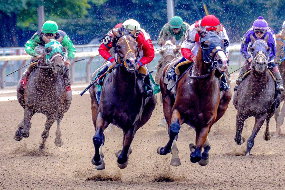
He began by acknowledging that Thoroughbred racing as a whole has made incredible strides in equine safety. However, he said that though racing injuries have dropped in recent years, they have since plateaued. He encouraged the industry to not “rest on their laurels” and noted that what could be viewed as “tolerance” of breakdowns in racing will soon be unacceptable to the public.
Forbes offered multiple ways in which racing can become more transparent—and therefore encourage its longevity. These include pre-race examinations that include both visual and hands-on, physical exams, and the use of advanced diagnostic imaging. Forbes acknowledges that there can be a discrepancy in exams between vets, including in their thoroughness and timing, so standardization of these tests would be necessary.
This lack of standardization between vets is one facet of a larger issue, which is lack of cohesion between racing jurisdictions. With different acceptable race-day medications, testing and protocols between tracks, it’s no wonder equine welfare is seen as subjective.
Here is where technology integration can play a main role in convincing the public that the racing industry cares deeply about their horses, alludes Forbes. He does note one caveat: the information these systems offer can be overwhelming. With so much data given by motion sensor technology, it can be difficult to parse out what is truly important. Forbes suggested the creation of well-thought-out matrix would help determine when a horse’s welfare is truly at risk; a culmination of issues would lead to a horse being “flagged” in a system, noting that it might be heading down a perilous path and measures could be taken to ensure its safety.
Another concern Forbes highlighted was the actual verbiage used regarding motion sensor technology: The ability to highlight a correlation between positive performance and motion sensor technology use, instead of its use being indicative that injury is imminent, would assist not only the public’s take on efforts made to ensure equine safety, but also encourage owners and trainers to embrace the technology. Though it may seem like simple semantics, the shift in focus would encourage many vets, owners and trainers – all of whom are charged with safeguarding equine welfare — to use motion sensor technologies.
“We must go forward and embrace change,” Forbes said. There is no silver bullet to solve all of racing’s issues, he noted. “We must work together as an industry” to save ourselves.
Solving the Challenges of Lameness Measurement in Racing
Equinosis’ Path Forward
By Andy Wolter, CEO
Dr. Forbes critique of the state of the art in inertial sensor measurement systems utility at the track highlights certain deficiencies the Equinosis team is focused on overcoming.
We understand the regimented schedule trainers and racehorses keep and limits of imposing additional activities – e.g. regular monitoring of lameness. Hence, we have been working to develop a measurement protocol for racehorses conducted with the jockey mounted and the horse jogging on the track.
The goals of this new protocol are:
- Full instrumentation in seconds.
- Complete data collection around the entire track from a stationary receiver near the track.
- Data collected while conducting a normal – not additional – activity (i.e. an exercise rider jogging the horse).

How inertial sensors meet the requirements at the track. Slide presented by Dr. Forbes.
What specifically are we doing to address the deficiencies Dr. Forbes noted?
| Requirements: |
Development Underway: |
Rapid Results
- Capturing and reporting data on hundreds of horses on a weekly basis.
Status: Instrumentation in ~ 2 minutes. Analysis within seconds, but data collected one horse at a time per system |
To reduce instrumentation time from 2 minutes to ~ 10 seconds. How?
- Eliminate the pastern sensor (to determine foot fall sequence, can be determined accurately with the pelvic mounted sensor). 2-sensor data collection option. 2022 release.
- Reposition the pelvic sensor to the saddle or saddle pad. Equivalence study underway. Estimated 2023 release.
- Attaching the sensors directly to saddle and bridle without tape or other disposables.
To enable concurrent data collection of multiple horses from one tablet computer. How?
- Next generation autonomous data collection sensor development underway. 2023 release.
|
Screen a Large Number of Horses
Status: Yes, but may require many Equinosis Q systems and labor to instrument horses, trot horses and operate tablet computers. |
To develop a collection protocol at a ridden jog, while employing the 2-sensor data collection option, with improved sensor attachment, and sensors capable of autonomous data collection.
- Ability for a single technician to keep up with instrumentation of many horses.
- No need for additional labor or activities to jog the horse in hand.
- Given multiple sets of autonomous sensors, many horses can be on the track simultaneously collecting data.
|
Interpretation is simple and unequivocal
Status: Designed for a veterinarian trained in its use. |
As the Q exists today, it is designed for a veterinarian skilled in equine moving evaluations who is trained in applying the data provided.
Certainly, we foresee a day where integrated Artificial Intelligence will assist data interpretation and expand the predictive capabilities at the veterinarian’s disposal horse-side. And we foresee in the very near future, the ability to support telemedicine to ensure appropriate veterinary expertise is available, regardless of where the data is collected or by whom.
Last year’s release of Lameness Locator 20/20 software with LL Intelligence and cloud interface was the first step down that path.
With the benefit of programmatic connectivity to the cloud, future data aggregation and large data set learning will be possible. In the future, expanded data analysis, information retrieval, and deeper learning via artificial intelligence will be possible horse-side.
However, we believe that any lameness diagnostic will require the interpretation by a skilled veterinarian. The biomechanics of equine lameness is not simple, nor is the clinical application of objective lameness data. |