


An Inside Look At Objective Evaluation: All-Access Investigation of the Equinosis Q
By Nancy S. Loving, DVM
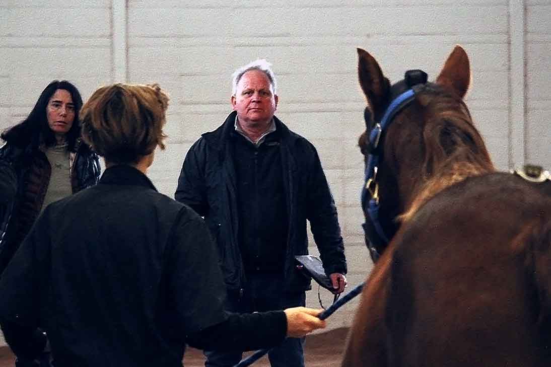
PICTURED ABOVE: Dr. Kevin Keagan (right) and Dr. Nancy Loving (left) watch as the horse is trotted on a straightaway.
FEB 2019 – As equine veterinarians, we are all too familiar with the challenges of diagnosing lameness and formulating a treatment plan. This winter I had a chance to learn hands on about a device that might change the whole paradigm of lameness evaluation. It’s called the Q with Lameness Locator, developed over the last three decades by Kevin Keegan, DVM, MS, DACVS at the University of Missouri. This device provides evidence-based, objective lameness measurement.
Admittedly, after performing three decades of lameness work that relied primarily on subjective evaluation, I was skeptical as to the value of the Q. I immersed myself in the educational videos by Equinosis – the company that manufactures and distributes the Q – before heading to Missouri. This information armed me with the fundamentals of how the device works to give context for forming my impressions of its value to equine practice.
I will share with you the basic premise of how the device works and then we’ll look at how well it worked when used on multiple cases during my visit to the University of Missouri veterinary teaching hospital.
Historical Context
Many may remember back to the mid 1980’s when the new technology available at that time centered on better pharmaceuticals to more reliably and safely manage pain and restraint – flunixin, Adequan, xylazine. Diagnostic ultrasound was just becoming available. It was a time when the personal computer was on the horizon as part of a home or business office, but still not yet a household word.
For veterinarians, recent decades of technologic advances have yielded amazing tools to aid in our quest to provide the best health care for our equine patients – ultrasound, digital radiology, thermal imaging, heart rate monitors, MRI, CT scan, to name a few. Computers are a seamless part of our lives – witness our pocket-sized cell phones, the Cloud for storage, powerful laptops that far exceed the capabilities of what NASA used to put a man on the moon.
No longer do equine practitioners need to rely solely on the single therapeutic plan of giving phenylbutazone and resting the horse because of an undefined lameness problem. The number of therapeutic options available is plentiful but success relies on an accurate diagnosis and appropriate therapeutic plan. Any tool available to accomplish this end is a bonus to equine practitioners, and this is where the Q has an important role.
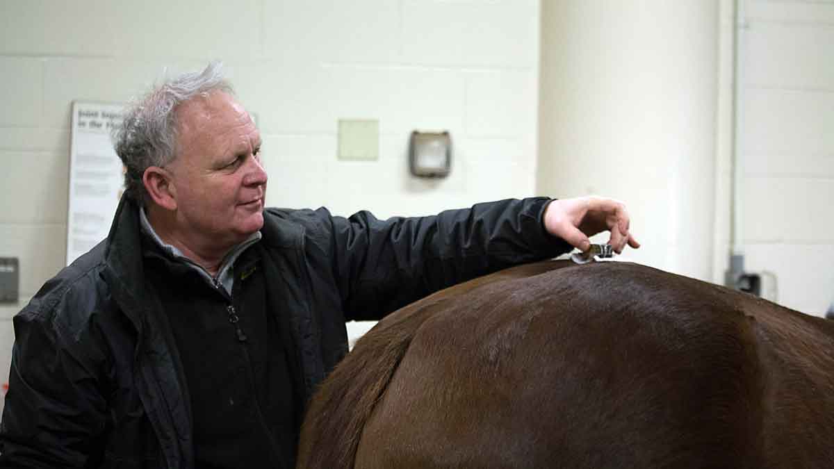
PICTURED: Dr. Kevin Keagan displays proper placement of the Q’s pelvic sensor.
The Equinosis Q with Lameness Locator
The Q with Lameness Locator is a sophisticated tool for detecting precision movement based on asymmetry in vertical acceleration of a horse’s torso at the trot. The basic premise of the device is that lameness – especially that which originates from pain – is caused by the decreased vertical force on the affected limb during stance. The algorithms of the input data obtained as a horse trots are translated into a plot that shows the veterinarian specifics about the amplitude and stride-by-stride variability, indicating decreased weight bearing and graphically represented with the timing in the stride cycle. Human athletes are able to talk to their doctors and explain what hurts. The Q serves a purpose as a communication device for the horse as he relays information to the computer with each step.
A report is generated that objectively pinpoints which limb or limbs are affected to cause a horse to exhibit the clinical sign of lameness. It takes only moments to place the inertial sensors on the horse’s head, pelvis, and right front pastern. The computer holds an antenna in its USB port that receives data wirelessly from as far away as 300 feet. The inertial sensors sample movement information ten times faster than the human eye. The results appear on the tablet screen almost instantly in what is referred to as a Q report, described below.
Using the Q can be a one-person operation in the field or clinic. The sensor information is broadcast to the computer tablet without the need for an assistant to aim something at the horse as it moves.
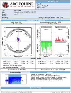
The Q Report
The Q report is the readout prepared on the tablet computer as a final analysis from the data collected by the inertial sensors as the horse trots. The information in the Q report gives precise measurements of differences in vertical head and pelvic movement between right and left halves of stride. There is also an AIDE report at the bottom of the page that synopsizes the data to note the degree of evidence of lameness for each limb (based on the stride by stride variability), the amplitude (from very mild to moderate/severe) of, and the timing (impact to push off) of lameness. Or, it might report that there is no evidence of lameness in a leg.
Not only is this helpful for diagnosing somewhat overt lameness, but the beauty of the tool is that it also reports on subtle lameness that may not be detectable to the naked eye. It also can be used as a prophylactic measure – veterinarians at one racing stable use it to monitor horses weekly for comparisons of each horse to itself. This facilitates recognition of subtle problems long before they simmer into something more troublesome.
In equine practice, this technique is useful for regular monitoring of a client’s horse before and during a competitive season. Or, it is useful to track progress during rehabilitation of an injury. Another use is as an objective tool when evaluating a horse for pre-purchase exam. For any movement evaluation, the Q is an excellent adjunctive device to diagnosis.
My Impressions
PICTURED RIGHT: Dr. Kevin Keagan (left) analyzing data generated by the Q with Dr. Nancy Loving (right).
Many practitioners pride themselves on having a keen eye for identifying a lame leg. Yet, there are always times when veterinarians are confounded by what they think they see. The Q takes the guesswork out of those situations and provides reliable, evidence-based information. As with any tool, it needs to be used intelligently, coupled with education on the correct procedures for instrumenting the horse and evaluating the data. Interpretation of results still depends on integrating clinical skills with data for best results. It is not practical to eliminate steps in a diagnostic workup just by looking at the Q report.
The device is sold only to veterinarians so that the scientific expertise of a veterinary education and clinical skillsets are used in conjunction with the diagnostic tool.
Clinical Cases
During the couple of days I spent with Dr. Keegan at the University of Missouri’s veterinary teaching hospital, we analyzed a number of horses for lameness exams using the Q.
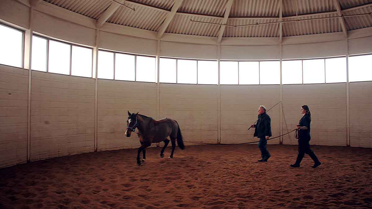
PICTURED LEFT: Dr. Keegan (left) instructs veterinary student Kaitlyn Galos (right) on proper techniques of a lunging trial.
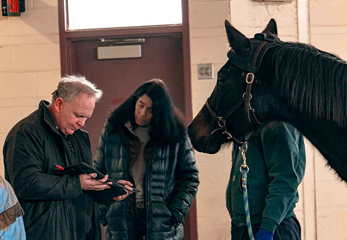
PICTURED: Dr. Kevin Keagan (left) stands with veterinary student Katelyn Galos (right) before starting the trial.
One of the cases we examined was the most telling about the value of the system. A Warmblood Event horse was trotted straightaway and on lunge circles, with no evident lameness to the naked eyes of multiple veterinarians. However, the data revealed a very mild lameness on the RF. With closer inspection, the horse resented repeated digital palpation of the proximal suspensory ligament. This led the practitioner to next take steps to image the ligament with diagnostic ultrasound.
Identifying a subclinical problem before it becomes more serious is an incredible aid to informing an owner about management strategies that help a horse stay sound and for veterinary intervention to proceed when appropriate. Identification of a suspensory ligament issue let the owner know that the horse needs rest and monitoring before returning him to rigorous work that could further injure the ligament. If we had only used the naked eye, it is possible that this beginning lameness would have gone undetected and the horse continued in work with the potential to develop a worse problem.
This one case made me think back to all the cursory “lameness checks” at clients’ farms pre- and during a competitive season and how easy it could have been to miss something like this. If the horse trots sound on lunge circles and the straightaway, then in the interest of time, it is often easy to forego a hands-on clinical evaluation of the limbs, thinking the horse is fine.
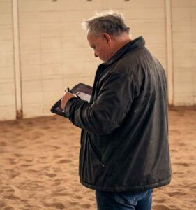
PICTURED : Dr. Kevin Keagan checking on trial data from the lunging trial being performed.
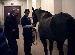
Another case involved a retired Event mare brought back for a recheck for a meniscal tear and an OCD lesion in a stifle. The objective was for the horse to be comfortable enough for a very young girl to ride the horse lightly. The mare provides emotional therapy in remarkable ways for the little girl and their bond is something special to see. First, the Q evaluated how well the previous treatment worked from the month before. The data plots from the previous and current visits were compared – there was improvement but more was desired. The diagnostic workup continued by injecting the stifle with anesthetic and triamcinolone to block the joint and treat it at the same time. Then, as expected, the stifle blocked sound but the Q picked up lameness in a front leg. The front limb lameness was addressed with further diagnostic workup. The young girl will have better success riding this horse once all lameness issues are sorted out and managed.
We also applied the inertial sensors to a horse with significant bilateral frontend lameness that was obvious even at a walk. The horse could be lunged on soft pack ground to obtain baseline information. Then nerve blocks were administered starting with a PDN block in the most lame forelimb. Once that leg blocked out well, the other leg received a PDN block. The owner had sent the horse to the clinic for neurectomy surgery. However, the Q revealed that not only was there severe bilateral lameness in the front end, but one limb didn’t respond to anesthesia until the horse received a high abaxial nerve block. In addition, a significant lameness in a rear limb was further identified. Taken at face value at the owner’s farm with only a subjective lameness assessment, this horse may have been put through an unnecessary surgery with poor results. While the obvious forelimb lameness issues were apparent to the naked eye, as the diagnostic workup proceeded, results from the Q report indicated another less obvious lameness and injury in the hind leg.
Sensor Placement
As my time at the University continued, we evaluated other horses by placing the inertial sensors correctly to obtain a Q report, and then the inertial sensors were put on incorrectly in a variety of configurations to test outcomes. The purpose of this mini study was to help devise new software that can recognize misplaced or misaligned sensors. The good news is that the RF pastern gyroscope can be turned nearly 90 degrees before the analysis is affected (the right front sensor determines the side and timing of measured lameness). The Equinosis team expects to implement what they learn from varying sensor placements by including that data in upcoming software this year.
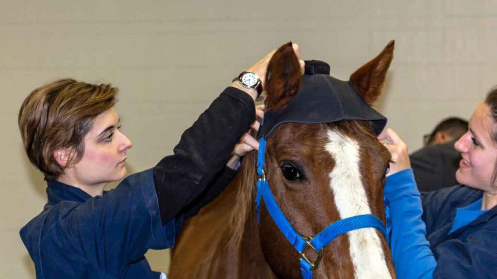
PICTURED: Veterinary students Eileen Larsen (left) and Katelyn Galos (right) equipping a horse with the Equinosis Q head bonnet.
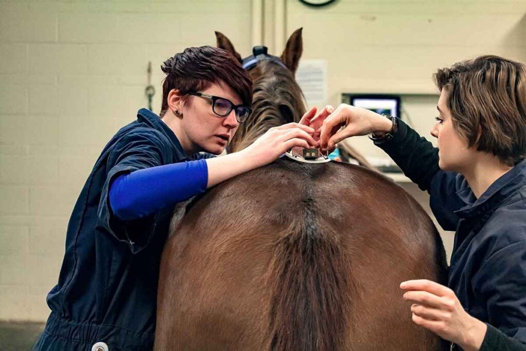
PICTURED: Veterinary students Rachael Schulte (left) and Eileen Larsen (right) ensuring the Q’s pelvic sensor is properly aligned.
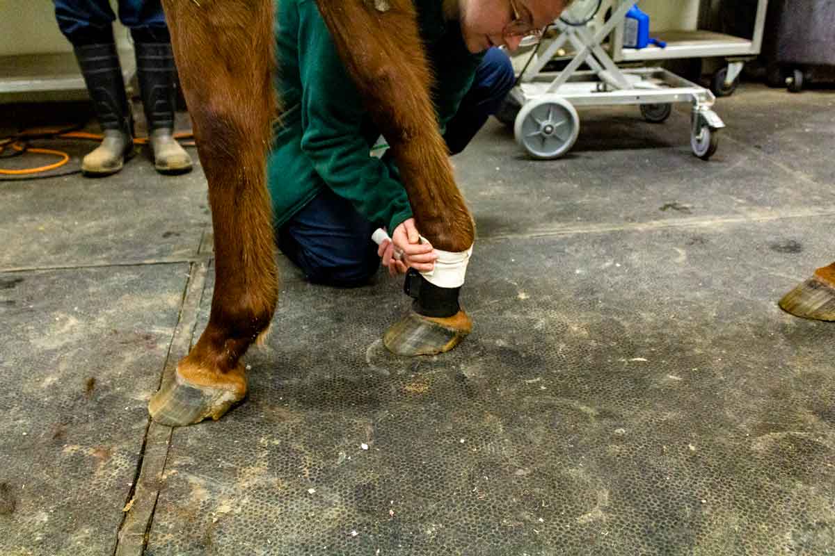
PICTURED: Student ensuring the right forelimb sensor is properly aligned and secured.
In Summary
My visit to Missouri was done with an open mind although with a little bit of skepticism. It turns out that I was pleasantly surprised by the consistency of the Q reports for each horse we looked at. Results of the Q aligned with further diagnostic workup that corroborated what the inertial sensors were reporting. A tool that more reliably identifies where to begin a diagnostic workup gives you a greater chance of achieving a definitive diagnosis. And, with that diagnosis you are able to provide therapy that works in the best interest of the horse.
Thinking back on the myriad of lameness workups done during my veterinary career, I wonder just how much better a lameness diagnostician I might have become with this device used as an adjunctive tool to my understanding. Dr. Keegan is to be applauded for his perseverance in developing this technology, and for his contribution to the equine industry.
***
Lo Jones
This is such a huge jump for the equine community. Your advanced services are known across the country and after reading this awesome article, you have far exceeded the equines expectations.